Human Stomach
Stomach
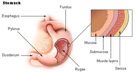
The major function of the stomach is to temporarily store food and release it slowly into the duodenum. It processes the food to a semi-solid chyme, which enables better contact with the mucous membrane of the intestine, thereby facilitating absorption of nutrients. In addition, the stomach is an important site of enzyme production.
The cardia surrounds the superior opening of the stomach. The rounded portion superior to the body and to the left of the cardia is the fundus. Inferior to the fundus is the large central portion of the stomach, called the body. The region of the stomach that connects to the duodenum is the pylorus. It has two parts, the pyloric antrum, which connects to the body of the stomach, and the pyloric canal, which leads into the duodenum. The pylorus communicates with the duodenum of the small intestine via the pyloric sphincter (valve). This valve regulates the passage of chyme from stomach to duodenum and it prevents backflow of chyme from duodenum to stomach (Tortora and Grabowski, 1996). The stomach wall is composed of four layers: mucosa, submucosa, muscularis and serosal.
The cardia surrounds the superior opening of the stomach. The rounded portion superior to the body and to the left of the cardia is the fundus. Inferior to the fundus is the large central portion of the stomach, called the body. The region of the stomach that connects to the duodenum is the pylorus. It has two parts, the pyloric antrum, which connects to the body of the stomach, and the pyloric canal, which leads into the duodenum. The pylorus communicates with the duodenum of the small intestine via the pyloric sphincter (valve). This valve regulates the passage of chyme from stomach to duodenum and it prevents backflow of chyme from duodenum to stomach (Tortora and Grabowski, 1996). The stomach wall is composed of four layers: mucosa, submucosa, muscularis and serosal.
Intestine Conditions
- Stomach flu (enteritis): Inflammation of the small intestine. Infections (from viruses, bacteria, or parasites) are the common cause.
- Small intestine cancer: Rarely, cancer may affect the small intestine. There are multiple types of small intestine cancer, causing about 1,100 deaths each year.
- Celiac disease: An "allergy" to gluten (a protein in most breads) causes the small intestine not to absorb nutrients properly. Abdominal pain and weight loss are usual symptoms.
- Carcinoid tumor: A benign or malignant growth in the small intestine. Diarrhea and skin flushing are the most common symptoms.
- Intestinal obstruction: A section of either the small or large bowel can become blocked or twisted or just stop working. Belly distension, pain, constipation, and vomiting are symptoms.
- Colitis: Inflammation of the colon. Inflammatory bowel disease or infections are the most common causes.
- Diverticulosis: Small weak areas in the colon's muscular wall allow the colon's lining to protrude through, forming tiny pouches called diverticuli. Diverticuli usually cause no problems, but can bleed or become inflamed.
- Diverticulitis: When diverticuli become inflamed or infected, diverticulitis results. Abdominal pain and constipation are common symptoms.
- Colon bleeding (hemorrhage): Multiple potential colon problems can cause bleeding. Rapid bleeding is visible in the stool, but very slow bleeding might not be.
- Inflammatory bowel disease: A name for either Crohn's disease or ulcerative colitis. Both conditions can cause colon inflammation (colitis).
- Crohn's disease: An inflammatory condition that usually affects the colon and intestines. Abdominal pain and diarrhea (which may be bloody) are symptoms.
- Ulcerative colitis: An inflammatory condition that usually affects the colon and rectum. Like Crohn's disease, bloody diarrhea is a common symptom of ulcerative colitis.
- Diarrhea: Stools that are frequent, loose, or watery are commonly called diarrhea. Most diarrhea is due to self-limited, mild infections of the colon or small intestine.
- Salmonellosis: Salmonella bacteria can contaminate food and infect the intestine. Salmonella causes diarrhea and stomach cramps, which usually resolve without treatment.
- Shigellosis: Shigella bacteria can contaminate food and infect the intestine. Symptoms include fever, stomach cramps, and diarrhea, which may be bloody.
- Traveler's diarrhea: Many different bacteria commonly contaminate water or food in developing countries. Loose stools, sometimes with nausea and fever, are symptoms.
- Colon polyps: Polyps are growths inside the colon. Colon cancer can often develop in these tumors after many years.
- Colon cancer: Cancer of the colon affects more than 100,000 Americans each year. Most colon cancer is preventable through regular screening.
- Rectal cancer: Colon and rectal cancer are similar in prognosis and treatment. Doctors often consider them together as colorectal cancer.
- Constipation: When bowel movements are infrequent or difficult.
- Irritable bowel syndrome (IBS): Irritable bowel syndrome, also known as IBS, is an intestinal disorder that causes irritable abdominal pain or discomfort, cramping or bloating, and diarrhea or constipation.
- Rectal prolapse: Part or all of the wall of the rectum can move out of position, sometimes coming out of the anus, when straining during a bowel movement.
- Intussusception: Occurring mostly in children, the small intestine can collapse into itself like a telescope. It can become life-threatening if not treated.
Small intestine
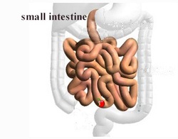
Small intestine is a bundled up, continuous tube located in your abdomen that receives food from the stomach at the duodenum, digests and absorbs food in the jejunum, and deposits food into the large intestine at the ileocecal valve. Stretched out, the small intestine of the average adult measures 5 meters in length and about 3 centimeters in diameter. Although, it is much longer than the large intestine, it has a much narrower diameter, thus accounting for its name. The majority of food digestion and nutrient absorption occurs in the small intestine.
The small intestine receives partially digested food from your stomach. At the point when food reaches the duodenum of the small intestine, it has been mixed with stomach acid and manually broken up by the contraction of the stomach walls. The smaller the pieces of food, the better able your body is to absorb the nutrients because many small pieces of food have much greater surface area than fewer large pieces of food. This surface area allows for greater exposure to the microvilli of the small intestine to absorb the maximum possible number of nutrients. Let’s examine how the body deals with carbohydrates, fats and proteins in the small intestine. Microvilli are small, finger like appendages attached to the larger villi that line the walls of your small intestine. Combined, the microvilli and villi increase the surface area inside your small intestine to 500 meters squared, whereas, if the intestinal walls were smooth with no ridges, the surface area would only be 1/2 a meter squared. Think of your microvilli as little nutrient vacuums inside your small intestine.
Fats
Lipid molecules (lipid means fat) must be broken down into very small globules in order to be taken up by the microvilli in your small intestine. Pancreatic lipase (“lip” refers to fat and “ase” means the “breaking down of”) is made in the pancreas and secreted through the pancreatic duct into the duodenum, the first portion of the small intestine after the stomach. Lipase is aided by bile from the gallbladder. Bile orients the fatty acids so that their hydrophobic (“water fearing”) heads point towards each other in the center, away from the watery intestinal walls, to allow the pancreatic lipase to break down triglycerides (fancy name for fat molecule) into glycerol and free fatty acids which can be absorbed by the microvilli.
Carbohydrates
Starchy and sugary foods consist mostly of carbohydrates. Carbohydrates are long strings of sugar molecules. Pancreatic amylase breaks down starch molecules into oligosaccharides. Then, sucrase, lactase and maltase (remember “ase” means the “breaking down of” and sucrose, lactose and maltose are different types of sugar molecules), break down the smaller components into molecules that can be absorbed and used in the body. It is interesting to note that lactase is absent in many adults; thus rendering them “lactose intolerant”. Lactose is the sugar in milk. Without lactase, our intestines are unable to digest milk products, resulting in a gaseous, upset, sick feeling, with diarrhea, and flatulence.
Protein
Protein digestion begins in the stomach with chemical and mechanical breakdown into smaller protein pieces and polypeptide chains. A protein is a long peptide chain (poly means “many”). Polypeptides are long chains of amino acids. Amino acids are absorbed by the microvilli of the small intestine. The pancreas secretes trypsin and chymotrypsin into the duodenum of the small intestine to break down proteins into amino acids.
How Does Absorption Happen?
The small intestine are networks of capillaries and lymph vessels called lacteals. Capillaries are the smallest components of your blood vessels, the site at which your blood exchanges nutrients, oxygen and waste products with your body’s tissues. Simply put, nutrients diffuse into your blood vessels through chemical and electrical gradients: in a very short, very oversimplified summary, substances will move from an area of high concentration to an area of low concentration and the cells in our tissues facilitate this by changing their chemical concentrations and electrical charges. Fat molecules are taken up by lacteals; little balloons of lymph tissue that merge with other lacteals to form lymphatic vessels responsible for circulating lymph fluid. Lacteals make it into our bloodstream for distribution to our tissues via the subclavian vein (sub = under and clavian = clavicle). Fats in the bloodstream are called chylomicrons.
Not all digested matter from our food makes it to the bloodstream. Whatever is left after our small intestine sucks out all of the nutrients passes onto our large intestine. Here, water and salt are reclaimed for the body, the waste is concentrated and comes out our anus as stool. What I find really interesting is that between the mouth and the anus is one, long continuous, open ended tube. What goes in, must come out. At risk of being too gross, I will mention that the color of your stool can be indicative of a number of illnesses of the pancreas, the liver, the gallbladder and more; since the color of stool is dependent both upon what we eat and what we use to digest food. for example, grey or whitish stool can indicate a problem with your liver or gallbladder.
Not all digested matter from our food makes it to the bloodstream. Whatever is left after our small intestine sucks out all of the nutrients passes onto our large intestine. Here, water and salt are reclaimed for the body, the waste is concentrated and comes out our anus as stool. What I find really interesting is that between the mouth and the anus is one, long continuous, open ended tube. What goes in, must come out. At risk of being too gross, I will mention that the color of your stool can be indicative of a number of illnesses of the pancreas, the liver, the gallbladder and more; since the color of stool is dependent both upon what we eat and what we use to digest food. for example, grey or whitish stool can indicate a problem with your liver or gallbladder.
Large Intestine
Front of abdomen, showing the large intestine, with the stomach and small intestine in gray outline.
Front of abdomen, showing surface markings for liver (red), and the stomach and large intestine (blue)
Latin intestinum crassum
Artery Superior mesenteric, Inferior mesenteric and Iliac arteries
Lymph inferior mesenteric lymph nodes
Dorlands/Elsevier Large intestine
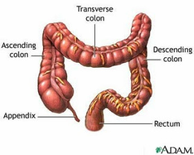
The large intestine (or “large bowel”) is the second-to-last part of the digestive system — the final stage of the alimentary canal is the anus — in vertebrate animals. Its function is to absorb water from the remaining indigestible food matter, and then to pass useless waste material from the body. This article is primarily about the human gut, though the information about its processes are directly applicable to most mammals.
The large intestine consists of the cecum and colon. It starts in the right iliac region of the pelvis, just at or below the right waist, where it is joined to the bottom end of the small intestine. From here it continues up the abdomen, then across the width of the abdominal cavity, and then it turns down, continuing to its endpoint at the anus.
The large intestine is about 1.5 metres (4.9 ft) long, which is about one-fifth of the whole length of the intestinal canal.
In Terminologia Anatomica the large intestine includes the cecum, colon, rectum, and anal canal. However, some sources exclude the anal canal.
Front of abdomen, showing surface markings for liver (red), and the stomach and large intestine (blue)
Latin intestinum crassum
Artery Superior mesenteric, Inferior mesenteric and Iliac arteries
Lymph inferior mesenteric lymph nodes
Dorlands/Elsevier Large intestine
The large intestine (or “large bowel”) is the second-to-last part of the digestive system — the final stage of the alimentary canal is the anus — in vertebrate animals. Its function is to absorb water from the remaining indigestible food matter, and then to pass useless waste material from the body. This article is primarily about the human gut, though the information about its processes are directly applicable to most mammals.
The large intestine consists of the cecum and colon. It starts in the right iliac region of the pelvis, just at or below the right waist, where it is joined to the bottom end of the small intestine. From here it continues up the abdomen, then across the width of the abdominal cavity, and then it turns down, continuing to its endpoint at the anus.
The large intestine is about 1.5 metres (4.9 ft) long, which is about one-fifth of the whole length of the intestinal canal.
In Terminologia Anatomica the large intestine includes the cecum, colon, rectum, and anal canal. However, some sources exclude the anal canal.
Disease And Disorders
- Gastroenteritis is an inflammation of the intestines. It is the most common disease of all the intestines.
- Ileus is a blockage of the intestines.
- Ileitis is an inflammation of the ileum.
- Colitis is an inflammation of the large intestine.
- Appendicitis is inflammation of the vermiform appendix located at the caecum. This is a potentially fatal disease if left untreated; most cases of appendicitis require surgical intervention.
- Angiodysplasia of the colon
- Chronic functional abdominal pain
- Colorectal cancer
- Constipation
- Diarrhea
- Hirschsprung's disease (aganglionosis)
- Intussusception
- Polyp (medicine)
- Pseudomembranous colitis
- Ulcerative colitis and toxic megacolon
How food is digested?
Most of the food is broken down and absorbed (taken in) into the blood where it is taken to all parts of the body. The food is used to give us energy to move, breathe, keep warm and many other activities. Food is also required to make us grow and to keep us healthy. Some of the food is not absorbed and is eventually lost from the body. This part of the food is eliminated from the body.
The mammalian digestive system is really a long tube which passes through the body. It starts at the mouth, where the food is ingested (taken in), and ends at the anus.
At the anus the parts of the food which are not useful are egested (moved out) of the body. When food is in this tube it is not really part of the body; it is just passing through it. It is something like travelling through the Alps by car. The car and its passengers are not part of the mountain but just passing through it. In the digestive system, the useful food is taken into the body. To take out the useful parts of the food, the digestive system breaks the food down into small pieces. This process is called digestion. The parts of the food which are not useful just pass through the body.
Mouth
The food is chewed in the mouth. We have already seen that different mammals have different kinds of teeth depending on the food they eat. As the food is chewed it is mixed with a liquid called saliva. We produce around one and a half cubic decimetres of saliva each day from special glands in our cheeks and under the tongue. When the food has been chewed enough, it is pushed to the back of the mouth by the tongue and swallowed.
Oesophagus
When the food is swallowed it moves from the back of the mouth into the first part of the digestive system, a tube called the oesophagus. The oesophagus moves the food through the chest region of the body which is called the thorax. Solid food passes through the oesophagus to the stomach in 5 seconds. Liquids only take 1 second to reach the stomach.
Stomach
The stomach is a part of the digestive system which is wider than the rest of the tube. It is shaped like a bag and it is used to store food. It is able to hold one and a half dm3 of food comfortably. The food stays in the stomach for about two and a half hours. While it is there it is mixed with juices, produced by the stomach wall, which break it down into much smaller pieces. Some of these pieces are so small that they start to dissolve in the juices. The semi-liquid food now passes into the small intestine.
Small Intestine
This part of the digestive system is a long, coiled tube, about 4 metres long. The food is mixed with more juices which come from the liver and the pancreas. These juices turn the food into even smaller pieces. A lot of the food is now so small that it is in solution. The useful parts of the dissolved food are absorbed (taken in) through the wall of the long tube. There are structures shaped like fingers which stick out from the wall of the small intestine. In the small intestine of a rat there are about 140 000 of these structures called villi (singular : villus). The villi increase the surface area of the gut wall so that more food can be absorbed. The parts of the food which are not absorbed stay in the tube and move into the next part of the digestive system, the large intestine.
Large Intestine
The tube which is called the large intestine is wider than the small intestine but not as long. It measures one and a half metres in length. Food can stay in the large intestine for a long time, up to 96 hours! It is here that a lot of the water contained in the food is absorbed into the body. This leaves the remaining parts of the food in a more solid condition.
Rectum and the Anus
The solid parts of the food which have not been absorbed are stored in the rectum. When the rectum is full, the remains of the food are passed out of the body through the anus.
All mammals have a similar digestive system but there are some differences. For example, there is a part of the digestive system which is called the appendix. It is found between the small intestine and the large intestine. The appendix is important in herbivores because it helps them digest plant food. Therefore it is quite large in an animal such as the rabbit. It is less important, however, in the human and is much smaller because of this. Sometimes a human appendix can be swollen and can cause a lot of pain. If this happens there has to be an operation to remove it. A person who has had his appendix taken out can continue to eat the same things as usual. This proves that the appendix is not essential in humans
