The Human Head
Human Head and Brain
The human head contains 22 bones, consisting the cranium and the facial bones. The cranium is formed by 8 bones: the frontal bone, two parietal bones, two temporal bones, the occipital bone in the back, the ethmoid bone behind the nose, and the sphenoid bone. The face consists of 14 bones including the maxilla (upper jaw) and mandible (lower jaw). (The skull has many little holes in its base which allow the cranial nerves to travel to their destinations.)
The cranium protects the brain, which, for an average adult male weighs about 1400 gram (49 oz). The brain of Russian novelist Turgenev, weighed 2021g (71 oz), Bismarck’s brain weighed 1807g (64 oz), while that of famous French statesman Gambetta was 1294g (46 oz). Female average brain mass is slightly less than that of males. The largest woman’s brain recorded weighed 1742g (61 oz). Einstein’s brain weighed 1230 gram (43.39 ounces), meaning Einstein’s brain was smaller than average.
Head Brain
 |
 |
Head and neck
Head and neck anatomy focuses on the structures of the head and neck of the human body, including the brain, bones, muscles, blood vessels, nerves, glands, nose, mouth, teeth, tongue, and throat. It is an area frequently studied in depth by surgeons, dentists, dental technicians, and speech language pathologists.
Musculoskeletal system
The head is positioned upon the superior portion of the vertebral column, attaching the skull upon C-1, (the atlas). The skeletal section of the head and neck forms the superior segment of the axial skeleton and comprises skull, hyoid bone, auditory ossicles, and cervical spine. The skull can be further subdivided into:
In a newborn, the junction of the parietal bones with the frontal and occipital bones, form the anterior (front) and posterior (back) fontanelle, or soft spots. The separation of the cranial bone plates at time of birth facilitate passage of the head of the fetus through the mother's birth canal, or pelvic girdle. The parietal bones, and occipital bone can overlap each other in the birth canal, and form the unusual looking "cone head" appearance in a newborn when delivered in a natural, or vaginal, delivery.
The occipital bone articulates with the atlas near the foramen magnum. The atlas articulates with the occipital condyle superiorly and the axis inferiorly. The spinal cord passes through the foramen magnum providing continuity for the central nervous system (CNS). Articulation of the neck includes: flexion, extension, (nodding yes), and rotation (shaking head no).
Circulatory system Blood circulates from the upper systemic loop originating at the aortic arch, and includes: the brachiocephalic artery, left common carotid and left subclavian artery. The head and neck are emptied of blood by the subclavian vein and jugular vein.
Blood supply
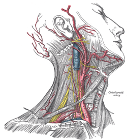
Right side of neck dissection showing the brachiocephalic, right common carotid artery and its branches
The brachiocephalic artery or trunk is the first and largest artery that branches to form the right common carotid artery and the right subclavian artery. This artery provides blood to the right upper chest, right arm, neck, and head, through a branch called right vertebral artery. The right and left vertebral artery feed into the basilar artery and upward to the Posterior cerebral artery, which provides most of the brain with oxygenated blood. The posterior cerebral artery and the posterior communicating artery are within the circle of Willis.
The left common carotid artery divides to form the: internal carotid artery (ICA) and an external carotid artery (ECA). The ICA supplies the brain. The ECA supplies the neck and face.
The left subclavian artery and the right subclavian artery, one on each side of the body form the internal thoracic artery, the vertebral artery, the thyrocervical trunk, and the costocervical trunk. The subclavian becomes the axiliary artery at the lateral border of the first rib. The left subclavian artery also provides blood to the left upper chest and left arm.
Blood-brain barrier
The Blood-brain barrier (BBB) is semi-permeable membrane that controls the capillary leak potential of the circulatory system. In most parts of the body, the smallest blood vessels, called capillaries, are lined with endothelial cells, which have small spaces between each individual cell so substances can move readily between the inside and the outside of the capillary. This is not the case in the brain. In the brain, the endothelial cells fit tightly together to create a tight junction and substances cannot pass out of the bloodstream.
Specialized glial cells called astrocytes form a tight junction or protective barrier around brain blood vessels and may be important in the development of the BBB. Astrocytes may be also be responsible for transporting ions (electrolytes) from the brain to the blood.
Blood return Blood from the brain and neck flows from: within the cranium via the internal jugular veins, a continuation of the sigmoid sinuses. The right and left external jugular veins drain from the parotid glands, facial muscles, scalp into the subclavian veins. The right and left vertebral veins drain the vertebrae and muscles into the right subclavian vein and into the superior vena cava, into the right atrium of the heart.
Musculoskeletal system
The head is positioned upon the superior portion of the vertebral column, attaching the skull upon C-1, (the atlas). The skeletal section of the head and neck forms the superior segment of the axial skeleton and comprises skull, hyoid bone, auditory ossicles, and cervical spine. The skull can be further subdivided into:
- (a) cranium, (8 bones: frontal, 2-parietal, occipital, 2-temporal, sphenoid, ethmoid), and
- (b) facial bones, (14 bones: 2-zygomatic, 2-maxillary, 2-palatine, 2-nasal, 2-lacrimal, vomer, 2-inferior conchae, mandible).
In a newborn, the junction of the parietal bones with the frontal and occipital bones, form the anterior (front) and posterior (back) fontanelle, or soft spots. The separation of the cranial bone plates at time of birth facilitate passage of the head of the fetus through the mother's birth canal, or pelvic girdle. The parietal bones, and occipital bone can overlap each other in the birth canal, and form the unusual looking "cone head" appearance in a newborn when delivered in a natural, or vaginal, delivery.
The occipital bone articulates with the atlas near the foramen magnum. The atlas articulates with the occipital condyle superiorly and the axis inferiorly. The spinal cord passes through the foramen magnum providing continuity for the central nervous system (CNS). Articulation of the neck includes: flexion, extension, (nodding yes), and rotation (shaking head no).
Circulatory system Blood circulates from the upper systemic loop originating at the aortic arch, and includes: the brachiocephalic artery, left common carotid and left subclavian artery. The head and neck are emptied of blood by the subclavian vein and jugular vein.
Blood supply
Right side of neck dissection showing the brachiocephalic, right common carotid artery and its branches
The brachiocephalic artery or trunk is the first and largest artery that branches to form the right common carotid artery and the right subclavian artery. This artery provides blood to the right upper chest, right arm, neck, and head, through a branch called right vertebral artery. The right and left vertebral artery feed into the basilar artery and upward to the Posterior cerebral artery, which provides most of the brain with oxygenated blood. The posterior cerebral artery and the posterior communicating artery are within the circle of Willis.
The left common carotid artery divides to form the: internal carotid artery (ICA) and an external carotid artery (ECA). The ICA supplies the brain. The ECA supplies the neck and face.
The left subclavian artery and the right subclavian artery, one on each side of the body form the internal thoracic artery, the vertebral artery, the thyrocervical trunk, and the costocervical trunk. The subclavian becomes the axiliary artery at the lateral border of the first rib. The left subclavian artery also provides blood to the left upper chest and left arm.
Blood-brain barrier
The Blood-brain barrier (BBB) is semi-permeable membrane that controls the capillary leak potential of the circulatory system. In most parts of the body, the smallest blood vessels, called capillaries, are lined with endothelial cells, which have small spaces between each individual cell so substances can move readily between the inside and the outside of the capillary. This is not the case in the brain. In the brain, the endothelial cells fit tightly together to create a tight junction and substances cannot pass out of the bloodstream.
Specialized glial cells called astrocytes form a tight junction or protective barrier around brain blood vessels and may be important in the development of the BBB. Astrocytes may be also be responsible for transporting ions (electrolytes) from the brain to the blood.
Blood return Blood from the brain and neck flows from: within the cranium via the internal jugular veins, a continuation of the sigmoid sinuses. The right and left external jugular veins drain from the parotid glands, facial muscles, scalp into the subclavian veins. The right and left vertebral veins drain the vertebrae and muscles into the right subclavian vein and into the superior vena cava, into the right atrium of the heart.
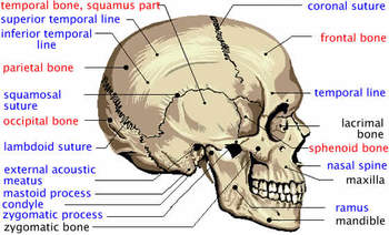
The Human Skull
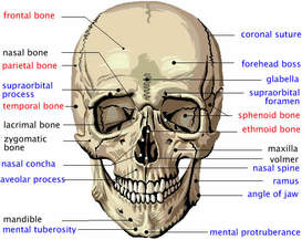
The human cranium and the facial bones are the foundation for the soft tissues of the face and head. Thus, much of the visible appearance of the human face depends upon the shapes and qualities of these bones. The cranium is that part of the skull that holds and protects the brain in a large cavity, called the cranial vault. Eight plate-like bones form the human cranium by fitting together at joints called sutures. The most important of these cranial bones for the appearance of the face is the frontal bone, which underlies the top of the face above the eyeballs. The human skull also includes 14 facial bones that form the lower front of the skull and provide the framework for most of the face that is important to psychological research. These 22 skull bones form other, smaller cavities besides the cranial vault, including those for the eyes, the internal ear, the nose, and the mouth. The important facial bones include the jaw bone or mandible, the maxilla or upper jaw, the zygomatic or cheek bone, and the nasal bone.
The shapes and features of the human skull determine much of the static appearances of the face and provide the basis for the features of physiognomy. Forensic pathologists and biologists can reconstruct the superficial appearance of a face merely from the human skull, as in the case of the Kennewick Man. The reconstruction of this skull revealed a facial appearance that indicates he is a descendant of a more ancient migration from Asia than that which brought the ancestors of the Indians (Amerinds), who settled widely in the Americas before the arrival of Europeans.
The skull bones are associated with many other features. Processes are areas where the bones have extra tissue to hold muscles and ligaments; lines are grooves in the bone from other developmental processes; foramina are holes in the bones through which nerves and blood vessels pass; sinuses are empty spaces in the bones that make the skull lighter. Some of these features affect the physiognomy of the face due to variations in thickness, size, location, and shape.
The diagrams below show the major external features of the human cranium and the major skull bones. The names in black are facial bones, those in red are cranial bones, and those in blue are features of the bones.
 |
 |
Head and Face
The face divided into six equal squares, two by three. The upper horizontal division is roughly at the 'third eye' level mid-forehead, the lower at the base of the nose. The eyes sit on the horizontal center, the mouth on the center of the lower third. The face may only be a combination of bone, muscles, and skin, but it is the body’s core means of expressing feelings and emotions.
Brain
The human brains consists of more than 100 billion neurons (nerve cells) through which the brain’s commands are sent in the form of electric pulses. These pulses travel at more than 400 km/h (250 mph), creating enough electricity to power a light bulb. The brain consumes more energy than any other organ, burning up a whopping 20% of the food we take in.
The left side of your brain controls the right side of your body and the right side of your brain controls the left side of your body.
It is estimated that the mental capacity of a 100-year old human with perfect memory could be represented by computer with 10 to the power of 15 bits (one petabit). At the current rate of computer chip development, that figure can be reached in about 35 years. However, that represents just memory capacity, not the extremely complex processes of thought creation and emotions.
Functions of the Brain
The human brain is a complex organ that allows us to think, move, feel, see, hear, taste, and smell. It controls our body, receives information, analyzes information, and stores information (our memories).
The brain produces electrical signals, which, together with chemical reactions, let the parts of the body communicate. Nerves send these signals throughout the body.
Size of the Human Brain
The average human brain weighs about 3 pounds (1300-1400 g).
At birth, the human brain weighs less than a pound (0.78-0.88 pounds or 350-400 g). As a child grows, the number of cell remains relatively stable, but the cells grow in size and the number of connections increases. The human brain reaches its full size at about 6 years of age.
Composition of the Brain
The brain consists of gray matter (40%) and white matter (60%) contained within the skull. Brain cells include neurons and glial cells.
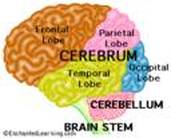
The brain has three main parts: the cerebrum, the cerebellum, and the brain stem (medulla).
Nourishment of the Brain
Although the brain is only 2% of the body's weight, it uses 20% of the oxygen supply and gets 20% of the blood flow. Blood vessels (arteries, capillaries, and veins) supply the brain with oxygen and nourishment, and take away wastes. If brain cells do not get oxygen for 3 to 5 minutes, they begin to die.
Cerebrospinal fluid (CSF) surrounds the brain.
The brain produces electrical signals, which, together with chemical reactions, let the parts of the body communicate. Nerves send these signals throughout the body.
Size of the Human Brain
The average human brain weighs about 3 pounds (1300-1400 g).
At birth, the human brain weighs less than a pound (0.78-0.88 pounds or 350-400 g). As a child grows, the number of cell remains relatively stable, but the cells grow in size and the number of connections increases. The human brain reaches its full size at about 6 years of age.
Composition of the Brain
The brain consists of gray matter (40%) and white matter (60%) contained within the skull. Brain cells include neurons and glial cells.
The brain has three main parts: the cerebrum, the cerebellum, and the brain stem (medulla).
Nourishment of the Brain
Although the brain is only 2% of the body's weight, it uses 20% of the oxygen supply and gets 20% of the blood flow. Blood vessels (arteries, capillaries, and veins) supply the brain with oxygen and nourishment, and take away wastes. If brain cells do not get oxygen for 3 to 5 minutes, they begin to die.
Cerebrospinal fluid (CSF) surrounds the brain.
Nervous System
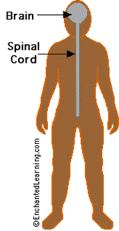
The brain and spinal cord make up the central nervous system (CNS). The brain is connected to the spinal cord, which runs from the neck to the hip area. The spinal cord carries nerve messages between the brain and the body.
The nerves that connect the CNS to the rest of the body are called the peripheral nervous system.
The autonomic nervous system controls our life support systems that we don't consciously control, like breathing, digesting food, blood circulation, etc.
Protection
The cells of the nervous system are quite fragile and need extensive protection from being crushed, being infected by disease organisms, and other harm. The brain and spinal cord are covered by a tough, translucent membrane, called the dura mater. Cerebration fluid (CSF) is a clear, watery liquid that surrounds the brain and spinal cord, and is also found throughout the ventricle (brain cavities and tunnels). CSF cushions the brain and spinal cord from jolts.
The cranium (the top of the skull) surrounds and protects the brain. The spinal cord is surrounded by vertebrae (hollow spinal bones). Also, some muscles serve to pad and support the spine.
More subtly, the blood-brain barrier protects the brain from chemical intrusion from the rest of the body. Blood flowing into the brain is filtered so that many harmful chemicals cannot enter the brain.
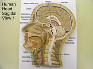
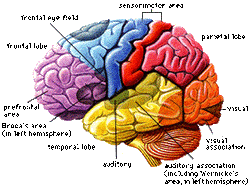
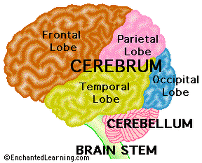
Structure Of Brain
The brain has three main parts, the cerebrum, the cerebellum, and the brain stem. The brain is divided into regions that control specific functions.
Cerebrum Cerebellum
|
Frontal Lobe
|
The Brain Stem
Hypothalamus
Optic Chiasm
Pituitary Gland
Spinal Cord
Pineal Body
Ventricles and Cerebral Aqueduct
 Structure And Functions Of Human Brain
|
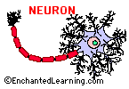
Brain Cells
The brain and spinal cord are made up of many cells, including neurons and glial cells. Neurons are cells that send and receive electro-chemical signals to and from the brain and nervous system. There are about 100 billion neurons in the brain. There are many more glial cells; they provide support functions for the neurons, and are far more numerous than neurons.
There are many type of neurons. They vary in size from 4 microns (.004 mm) to 100 microns (.1 mm) in diameter. Their length varies from a fraction of an inch to several feet.
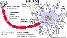
Neurons are nerve cells that transmit nerve signals to and from the brain at up to 200 mph. The neuron consists of a cell body (or soma) with branching dendrites (signal receivers) and a projection called an axon, which conduct the nerve signal. At the other end of the axon, the axon terminals transmit the electro-chemical signal across a synapse (the gap between the axon terminal and the receiving cell). The word "neuron" was coined by the German scientist Heinrich Wilhelm Gottfried von Waldeyer-Hartz in 1891 (he also coined the term "chromosome").
The axon, a long extension of a nerve cell, and take information away from the cell body. Bundles of axons are known as nerves or, within the CNS (central nervous system), as nerve tracts or pathways. Dendrites bring information to the cell body.
Myelin coats and insulates the axon (except for periodic breaks called nodes of Ranvier), increasing transmission speed along the axon. Myelin is manufactured by Schwann's cells, and consists of 70-80% lipids (fat) and 20-30% protein.
The cell body (soma) contains the neuron's nucleus (with DNA and typical nuclear organelles). Dendrites branch from the cell body and receive messages.
A typical neuron has about 1,000 to 10,000 synapses (that is, it communicates with 1,000-10,000 other neurons, muscle cells, glands, etc.).
Different Type of Neurons
There are different types of neurons. They all carry electro-chemical nerve signals, but differ in structure (the number of processes, or axons, emanating from the cell body) and are found in different parts of the body.
Life Span of Neuron
Unlike most other cells, neurons cannot regrow after damage (except neurons from the hippocampus). Fortunately, there are about 100 billion neurons in the brain.
Glial Cells
Glial cells make up 90 percent of the brain's cells. Glial cells are nerve cells that don't carry nerve impulses. The various glial (meaning "glue") cells perform many important functions, including: digestion of parts of dead neurons, manufacturing myelin for neurons, providing physical and nutritional support for neurons, and more. Types of glial cells include Schwann's Cells, Satellite Cells, Microglia, Oligodendroglia, and Astroglia.
Neuroglia (meaning "nerve glue") are the another type of brain cell. These cells guide neurons during fetal development.
There are many type of neurons. They vary in size from 4 microns (.004 mm) to 100 microns (.1 mm) in diameter. Their length varies from a fraction of an inch to several feet.
Neurons are nerve cells that transmit nerve signals to and from the brain at up to 200 mph. The neuron consists of a cell body (or soma) with branching dendrites (signal receivers) and a projection called an axon, which conduct the nerve signal. At the other end of the axon, the axon terminals transmit the electro-chemical signal across a synapse (the gap between the axon terminal and the receiving cell). The word "neuron" was coined by the German scientist Heinrich Wilhelm Gottfried von Waldeyer-Hartz in 1891 (he also coined the term "chromosome").
The axon, a long extension of a nerve cell, and take information away from the cell body. Bundles of axons are known as nerves or, within the CNS (central nervous system), as nerve tracts or pathways. Dendrites bring information to the cell body.
Myelin coats and insulates the axon (except for periodic breaks called nodes of Ranvier), increasing transmission speed along the axon. Myelin is manufactured by Schwann's cells, and consists of 70-80% lipids (fat) and 20-30% protein.
The cell body (soma) contains the neuron's nucleus (with DNA and typical nuclear organelles). Dendrites branch from the cell body and receive messages.
A typical neuron has about 1,000 to 10,000 synapses (that is, it communicates with 1,000-10,000 other neurons, muscle cells, glands, etc.).
Different Type of Neurons
There are different types of neurons. They all carry electro-chemical nerve signals, but differ in structure (the number of processes, or axons, emanating from the cell body) and are found in different parts of the body.
- Sensory neurons or Bipolar neurons carry messages from the body's sense receptors (eyes, ears, etc.) to the CNS. These neurons have two processes. Sensory neuron account for 0.9% of all neurons. (Examples are retinal cells, olfactory epithelium cells.)
- Motoneurons or Multipolar neurons carry signals from the CNS to the muscles and glands. These neurons have many processes originating from the cell body. Motoneurons account for 9% of all neurons. (Examples are spinal motor neurons, pyramidal neurons, Purkinje cells.)
- Interneurons or Pseudopolare (Spelling) cells form all the neural wiring within the CNS.These have two axons (instead of an axon and a dendrite). One axon communicates with the spinal cord; one with either the skin or muscle. These neurons have two processes. (Examples are dorsal root ganglia cells).
Life Span of Neuron
Unlike most other cells, neurons cannot regrow after damage (except neurons from the hippocampus). Fortunately, there are about 100 billion neurons in the brain.
Glial Cells
Glial cells make up 90 percent of the brain's cells. Glial cells are nerve cells that don't carry nerve impulses. The various glial (meaning "glue") cells perform many important functions, including: digestion of parts of dead neurons, manufacturing myelin for neurons, providing physical and nutritional support for neurons, and more. Types of glial cells include Schwann's Cells, Satellite Cells, Microglia, Oligodendroglia, and Astroglia.
Neuroglia (meaning "nerve glue") are the another type of brain cell. These cells guide neurons during fetal development.
