Human Nervous System
Nerves
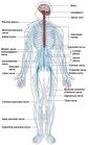
The nerves serve as paths for the transmission of nerve impulses along axons. Nerves are found only in the peripheral nervous system. Depending on the direction of the signals they conduct, they are classified into afferent and efferent nerves. The afferent ones conduct signals from sensory neurons to the central nervous system, while the efferent ones conduct signals from the central nervous system along motor neurons to muscles or glands. There are some nerves which can function like both afferent and efferent ones. They are called mixed nerves. Let's look at the major nerves given in the human nervous system diagram below.
Musculocutaneous Nerve : It is a part of the brachial plexus. It runs through the neck, the armpit area and ends in the arm. It serves the bicep muscles and the skin of the forearm.
Radial Nerve : It is also a part of the brachial plexus. It supplies the triceps brachii muscle of the arm and a part of the forearm along with its associated joints and skin.
Median Nerve : It is one of the main nerves that originate from the brachial plexus. It runs down the arm and enters the forearm. The median nerve is the only nerve passing through the carpal tunnel.
Ilio-hypogastric Nerve : It originates from the first lumbar nerve and supplies the abdominal muscles along with skin of some parts of the abdominal wall.
Obturator Nerve : It is a mixed nerve that arises from the lumbar plexus. It supplies the adductor, gracilis and obturator externus muscles. It also supplies a part of the skin of the thigh, hip and knee joints.
Genitofemoral Nerve : It arises in the lumbar plexus and bifurcates into two branches, namely, genital and femoral. Its branches run through the skin of the scrotum and to the upper anterior aspect of the thigh.
Ulnar Nerve : It runs near the ulna bone and is directly connected to the little finger and half of the ring finger. It supplies the tips of these fingers and the far back of the fingernail beds. It is the largest nerve which is unprotected by muscle or bone.
Common Peroneal Nerve : Also known as the common fibular nerve, it is half the size of the tibial nerve and originates from the branches of the lumbar and sacral nerves. It runs obliquely along the side of the depression at the back of the knee joint to the head of the calf bone.
Deep Peroneal Nerve : Also known as the deep fibular nerve, it originates at the bifurcation of the common peroneal nerve, comes above the middle of the leg and then to the front of the ankle joint. The deep peroneal nerve supplies muscular branches to some parts of the leg and the ankle joint.
Superficial Peroneal Nerve : It supplies the peroneus longus, a muscle in the lateral compartment of the leg and the peroneus brevis, a smaller muscle lying under the peroneus longus. This nerve supplies musclular branches to the longus and the brevis muscles and cutaneous filaments to the skin of the lower part of the leg.
Tibial Nerve : The tibial nerve is a branch of the sciatic nerve. It passes through the depression at the back of the knee joint, where it gives of an articular branch to the knee joint and a cutaneous branch that becomes the sural nerve
Saphenous Nerve : It is the largest cut aneous branch of the femoral nerve. It supplies cutaneous branches to the skin of the leg and foot in the region between the knee and the ankle.
Sciatic Nerve : Also known as the is chiatic nerve, the sciatic nerve is a nerve fiber that begins in the lower back and ends in the lower limb. It supplies the skin of the leg and the muscles of the leg, foot and back of the thigh.
Pudental Nerve : Originating in the sacral region of the spinal cord, it is formed from the second, third and fourth sacral nerves. It is located in the pelvic region and it supplies the external genitalia of both men and women.
Femoral Nerve : It is located inside the leg and supplies muscles that help in bending and straightening the leg. It is the largest branch of the lumbar plexus.
Subcostal Nerve : It is the vertical branch of the 12th thoracic nerve and supplies some parts of the abdominal muscles. It supplies branches to the skin of the lower abdominal wall and the gluteal region. It passes along the border of the 12th rib.
Intercostal Nerves : The ventral branches of the thoracic nerves are known as intercostal nerves. The first two intercostal nerves supply fibers to the upper limb, the next four, to the thorax and the lower five to the thorax and abdomen.
Nervous System
The nervous system of vertebrate human (including animals) is divided into the central nervous system (CNS) and peripheral nervous system (PNS). The central nervous system (CNS) is the largest part, and includes the brain and spinal cord. The spinal cavity contains the spinal cord, while the head contains the brain. The CNS is enclosed and protected by meanings, a three-layered system of membranes, including a tough, leathery outer layer called the dura mater. The brain is also protected by the skull and the spinal cord by the vertebrae.
The peripheral nervous system (PNS) is a collective term for the nervous system structures that do not lie within the CNS. The large majority of the axon bundles called nerves are considered to belong to the PNS, even when the cell bodies of the neurons to which they belong reside within the brain or spinal cord. The PNS is divided into somatic and visceral parts. The somatic part consists of the nerves that innervate the skin, joints, and muscles. The cell bodies of somatic sensory neurons lie in dorsal root ganglia of the spinal cord. The visceral part, also known as the autonomic nervous system, contains neurons that innervate the internal organs, blood vessels, and glands. The autonomic nervous system itself consists of two parts:
Horizontal bisection of the head of an adult man, showing skin, skull, and brain with grey matter (brown in this image) and underlying white matter.
The vertebrate nervous system can also be divided into areas called grey matter ("gray matter" in American spelling) and white matter. Grey matter (which is only grey in preserved tissue, and is better described as pink or light brown in living tissue) contains a high proportion of cell bodies of neurons.
White matter is composed mainly of myelinated axons, and takes its color from the myelin. White matter includes all of the peripheral nerves, and much of the interior of the brain and spinal cord. Grey matter is found in clusters of neurons in the brain and spinal cord, and in cortical layers that line their surfaces.
There is an anatomical convention that a cluster of neurons in the brain or spinal cord is called a nucleus, whereas a cluster of neurons in the periphery is called a ganglion. There are, however, a few exceptions to this rule, notably including the part of the fore-brain called the basal gangli.
The peripheral nervous system (PNS) is a collective term for the nervous system structures that do not lie within the CNS. The large majority of the axon bundles called nerves are considered to belong to the PNS, even when the cell bodies of the neurons to which they belong reside within the brain or spinal cord. The PNS is divided into somatic and visceral parts. The somatic part consists of the nerves that innervate the skin, joints, and muscles. The cell bodies of somatic sensory neurons lie in dorsal root ganglia of the spinal cord. The visceral part, also known as the autonomic nervous system, contains neurons that innervate the internal organs, blood vessels, and glands. The autonomic nervous system itself consists of two parts:
- Sympathetic nervous system
- Parasympathetic nervous system.
Horizontal bisection of the head of an adult man, showing skin, skull, and brain with grey matter (brown in this image) and underlying white matter.
The vertebrate nervous system can also be divided into areas called grey matter ("gray matter" in American spelling) and white matter. Grey matter (which is only grey in preserved tissue, and is better described as pink or light brown in living tissue) contains a high proportion of cell bodies of neurons.
White matter is composed mainly of myelinated axons, and takes its color from the myelin. White matter includes all of the peripheral nerves, and much of the interior of the brain and spinal cord. Grey matter is found in clusters of neurons in the brain and spinal cord, and in cortical layers that line their surfaces.
There is an anatomical convention that a cluster of neurons in the brain or spinal cord is called a nucleus, whereas a cluster of neurons in the periphery is called a ganglion. There are, however, a few exceptions to this rule, notably including the part of the fore-brain called the basal gangli.
What is the nervous system?
The nervous system is a very important part of the human body. It is in charge of all of the actions and descions you make through-out the day. Its main conductor is your spinal cord. It is also made up of many other parts of your body incuding your brain and other many small cells found everywhere in your body. All of these parts have different functions and heave a variety of things they do for you through-out the day and your life.The nervous system is a highly specialized system whose main parts are nerves called neurons.
What is the part of nervous system?
There are many parts to the nervous system. The main parts of it that are most commonly known, are the brain, the spinal cord, and neurons. Some other parts of the nervous system include the 1st cervical nerve, 1st lumbar nerve, 1st sacral nerve, and 1st thoracic nerve. It also includes many facial nerves and femoral nerves. Every single part of the nervous system controls how you think, feel, and act. It makes up who you are and without it going to work everyday you would not be able to do anything.
Mode of Control of Nervous System:
There are two control systems in the human body which control and coordinate the body functions: nervous system and hormonal system. Nervous control differs from the hormonal control in following characteristics:
Basic Functions of Nervous System:
1. It controls and coordinates the body functions.
2. It receives sensory impulses from the sensory organs through afferent nerve fibers (sensory function), analyzes and interprets this information and initiates the motor impulses which are carried to effectors organs like muscles and glands to show response (motor function).
3. It keeps the previous stimuli as the experiences or memory which guide the animal in future.
4. It coordinates the visceral functions to maintain a homeostasis in the body fluids.
What is the part of nervous system?
There are many parts to the nervous system. The main parts of it that are most commonly known, are the brain, the spinal cord, and neurons. Some other parts of the nervous system include the 1st cervical nerve, 1st lumbar nerve, 1st sacral nerve, and 1st thoracic nerve. It also includes many facial nerves and femoral nerves. Every single part of the nervous system controls how you think, feel, and act. It makes up who you are and without it going to work everyday you would not be able to do anything.
Mode of Control of Nervous System:
There are two control systems in the human body which control and coordinate the body functions: nervous system and hormonal system. Nervous control differs from the hormonal control in following characteristics:
- The nervous control is rapid.
- It takes place through electrical signals called nerve impulses.
- Nerve impulses travel in specific direction
Basic Functions of Nervous System:
1. It controls and coordinates the body functions.
2. It receives sensory impulses from the sensory organs through afferent nerve fibers (sensory function), analyzes and interprets this information and initiates the motor impulses which are carried to effectors organs like muscles and glands to show response (motor function).
3. It keeps the previous stimuli as the experiences or memory which guide the animal in future.
4. It coordinates the visceral functions to maintain a homeostasis in the body fluids.
Nervous cells
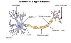
The nervous system is primarily made up of two categories of cells: neurons and cells. The nervous system is defined by the presence of a special type of cell—the neuron (sometimes called "neurone" or "nerve cell").
Neurons
Neuron can be considered as the basic unit of the nervous system, which processes and transmits information by means of electrochemical signals. Sensory neurons respond to external stimuli that affect the sensory organ cells. Motor neurons, on receiving signals from the central nervous system, bring about responses at the target organs. Interneurons act as the connectors between neurons. Neurons are of different shapes and sizes and their complex interconnections add to the complexity of the nervous system. The complexity is pretty evident from the human nervous system diagram given above. The human brain contains 86.1 billion neurons.
Glial
Glial or glial cells, as they are called, are non-neural cells that play a vital role in maintaining homeostasis and protecting the brain's neurons. The glial cells surround the neurons to hold them in place, supply them with oxygen and nutrients, isolate the neurons from one another and remove dead neurons. The human brain contains about 84.6 billion glial; that's almost equal to the number of neurons it contains.
The human nervous system can be divided into two parts, central and peripheral. The central nervous system (CNS) consists of the brain and the spinal cord, while the peripheral nervous system (PNS) consists of sensory neurons, ganglia (clusters of neurons) and nerves. Here is a labeled human nervous system diagram that you can refer to, while you read about the human nervous system function and parts.
Central nervous system
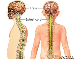
The central nervous system coordinates the functioning of all parts of the human body and is the largest part of the nervous system. It is enveloped by a set of membranes known as meninges that protect the brain and the spinal cord. They also have their own protective covers! The skull protects the brain while the vertebrae and spinal cavity shield the delicate spinal cord. To be precise, the brain is placed in the cranial cavity and the spinal cord in the spinal cavity. Let's take a closer look at the central nervous system parts and functions.
Brain
The brain is the center of the human nervous system and is a highly complex organ. Even a braniac won't find it very easy to understand the brain! The human brain is about three times larger than the brain of a typical mammal.
The brain can be said to have three main parts, the brain stem, the cerebrum and the cerebellum. The cerebrum is associated with information storage and processing; the cerebellum is responsible for balance, posture and coordination of movements; and the brain stem plays a vital role in controlling breathing and heart rate along with some other important body processes. Along with the skull, the brain is also protected by the cerebrospinal fluid in which it is suspended. It's strange yet true that the brain floats in a fluid!
For a detailed study of brain anatomy and its functions,
Spinal Cord
The spinal cord is a long tubular structure composed of nervous tissue and support cells. It is around 45 cm long in men and 43 cm long in women. It extends from the brain up to the space between the first and the second lumbar vertebrae. It transmits neural signals between the brain and other body parts. It is the spinal cord which connects the brain and the peripheral nervous system.
More on
The spinal nerves arise from the spinal column. The top section of the spine is the cervical section, which contains nerves that innervate muscles of the head, neck and thoracic cavity, as well as transmit sensory information to the CNS.
The cervical spine section contains seven vertabrae, C-1 through C-7, and eight nerve pairs, C-1 through C-8.
There is the formation of an extensive network of nerve groups or tracts attaching to the spinal cord in arrangements called rami or plexus.
The sensory branches of spinal nerves include: lesser occipital, C-2, great auricular, (C-2 and C-3); transverse cervical, C-2 and C-3; and supraclavicular, C-3 and C-4. These nerve groups transmit afferent (sensory) information from the scalp, neck, and shoulders to the brain.
The motor branches of spinal nerves include: ansa cervicalis, dividing into a superior root, C-1, and an inferior root, C-2 and C-3, and the phrenic nerve, C-3 to C-5, the segmental nerve branches, C-1 to C-5. These nerve groups transmit efferent nerve (motor) information from the brain to muscle groups of the scalp, neck, diaphragm (anatomy), and shoulders.
Additionally there are: (C5-C8, and T1) Brachial plexus, providing the entire nerve supply of the shoulder and upper limb; and includes supraclavicular branches (dorsal scapular, suprascapular, long thoracic) lateral cord (musculocutaneous, lateral antibrachial cutaneous, lateral head of median nerve), medial cord (ulnar, medial head of median nerve, medial antibrachial cutaneous, medial brachial cutaneous), posterior cord (axillary, radial), controlling the arm.
Damage to a person's spinal cord above C-5 may result in respiratory arrest and death if medicinal aid does not intervene.
Endocrine system The endocrine system is under the direct supervision of the nervous system, using the negative feedback principal of homeostasis, to create hormones which act as chemical instant messengers. The hypothalamus connects directly to the pituitary gland, both through the circulatory system and by direct connection of neurons. Also, within the cranium, the pineal gland, which attaches to the thalamus, controls the body's 24 hour rhythms circadian rhythm through the release of melatonin. Endocrine indicates that the secretion is used within the body. Endocrine glands are termed as ductless and release their secretions directly into the blood.
pituitary gland
The pituitary gland is also called hypophysis, or master gland. It secretes hormones that directly impact the body as well as hormones that indirectly control body functions because they activate other endocrine glands, such as the adrenal cortex (ACTH) and the thyroid gland (TSH). These two glands when stimulated by pituitary hormones then release their own hormones. The pituitary gland has two lobes, the anterior lobe and the posterior lobe. The anterior lobe secretes: growth hormone (GH), Luteinizing hormone (LH), Follicle stimulating hormone (FSH), Adrenocorticotropic hormone (ACTH), Thyroid-stimulating hormone (TSH), Prolactin (PRL), and the posterior lobe secretes: Antidieuretic hormone (ADH), and Oxytocin (OT). There is an intermediate lobe, in adult humans it is just a thin layer of cells between the anterior and posterior pituitary, nearly indistinguishable from the anterior lobe. The intermediate lobe produces melanocyte-stimulating hormone (MSH).
Thyroid glands
In the neck are the thyroid and parathyroid glands, that secrete hormones that control metabolism and blood calcium levels. The four parathyroid glands are situated upon the dorsal (back) surface of the thyroid gland.
Brain
The brain is the center of the human nervous system and is a highly complex organ. Even a braniac won't find it very easy to understand the brain! The human brain is about three times larger than the brain of a typical mammal.
The brain can be said to have three main parts, the brain stem, the cerebrum and the cerebellum. The cerebrum is associated with information storage and processing; the cerebellum is responsible for balance, posture and coordination of movements; and the brain stem plays a vital role in controlling breathing and heart rate along with some other important body processes. Along with the skull, the brain is also protected by the cerebrospinal fluid in which it is suspended. It's strange yet true that the brain floats in a fluid!
For a detailed study of brain anatomy and its functions,
- Diagram of the Brain and its Functions
- Left Brain Functions
- Right Brain Functions
Spinal Cord
The spinal cord is a long tubular structure composed of nervous tissue and support cells. It is around 45 cm long in men and 43 cm long in women. It extends from the brain up to the space between the first and the second lumbar vertebrae. It transmits neural signals between the brain and other body parts. It is the spinal cord which connects the brain and the peripheral nervous system.
More on
- Anatomy of Central Nervous System
- Central Nervous System Disorders
The spinal nerves arise from the spinal column. The top section of the spine is the cervical section, which contains nerves that innervate muscles of the head, neck and thoracic cavity, as well as transmit sensory information to the CNS.
The cervical spine section contains seven vertabrae, C-1 through C-7, and eight nerve pairs, C-1 through C-8.
There is the formation of an extensive network of nerve groups or tracts attaching to the spinal cord in arrangements called rami or plexus.
The sensory branches of spinal nerves include: lesser occipital, C-2, great auricular, (C-2 and C-3); transverse cervical, C-2 and C-3; and supraclavicular, C-3 and C-4. These nerve groups transmit afferent (sensory) information from the scalp, neck, and shoulders to the brain.
The motor branches of spinal nerves include: ansa cervicalis, dividing into a superior root, C-1, and an inferior root, C-2 and C-3, and the phrenic nerve, C-3 to C-5, the segmental nerve branches, C-1 to C-5. These nerve groups transmit efferent nerve (motor) information from the brain to muscle groups of the scalp, neck, diaphragm (anatomy), and shoulders.
Additionally there are: (C5-C8, and T1) Brachial plexus, providing the entire nerve supply of the shoulder and upper limb; and includes supraclavicular branches (dorsal scapular, suprascapular, long thoracic) lateral cord (musculocutaneous, lateral antibrachial cutaneous, lateral head of median nerve), medial cord (ulnar, medial head of median nerve, medial antibrachial cutaneous, medial brachial cutaneous), posterior cord (axillary, radial), controlling the arm.
Damage to a person's spinal cord above C-5 may result in respiratory arrest and death if medicinal aid does not intervene.
Endocrine system The endocrine system is under the direct supervision of the nervous system, using the negative feedback principal of homeostasis, to create hormones which act as chemical instant messengers. The hypothalamus connects directly to the pituitary gland, both through the circulatory system and by direct connection of neurons. Also, within the cranium, the pineal gland, which attaches to the thalamus, controls the body's 24 hour rhythms circadian rhythm through the release of melatonin. Endocrine indicates that the secretion is used within the body. Endocrine glands are termed as ductless and release their secretions directly into the blood.
pituitary gland
The pituitary gland is also called hypophysis, or master gland. It secretes hormones that directly impact the body as well as hormones that indirectly control body functions because they activate other endocrine glands, such as the adrenal cortex (ACTH) and the thyroid gland (TSH). These two glands when stimulated by pituitary hormones then release their own hormones. The pituitary gland has two lobes, the anterior lobe and the posterior lobe. The anterior lobe secretes: growth hormone (GH), Luteinizing hormone (LH), Follicle stimulating hormone (FSH), Adrenocorticotropic hormone (ACTH), Thyroid-stimulating hormone (TSH), Prolactin (PRL), and the posterior lobe secretes: Antidieuretic hormone (ADH), and Oxytocin (OT). There is an intermediate lobe, in adult humans it is just a thin layer of cells between the anterior and posterior pituitary, nearly indistinguishable from the anterior lobe. The intermediate lobe produces melanocyte-stimulating hormone (MSH).
Thyroid glands
In the neck are the thyroid and parathyroid glands, that secrete hormones that control metabolism and blood calcium levels. The four parathyroid glands are situated upon the dorsal (back) surface of the thyroid gland.
Peripheral Nervous System
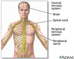
The central nervous system communicate with the other body organs? It is through the peripheral nervous system. Functionally, the peripheral nervous system can be divided into two parts; the somatic nervous system and the autonomic nervous system, the somatic nervous system is responsible for bodily activities that are under conscious control. For example, controlling body movements and receiving external stimuli. The autonomic nervous system is further divided into sympathetic, parasympathetic and enteric nervous systems. The sympathetic nervous system governs the bodily responses to impending dangers, while the parasympathetic system is responsible for bodily actions that help in relaxation of body organs that are hyperctive. The enteric nervous system specifically manages the functioning of the digestive system. Let's take a close look at what constitutes the peripheral nervous system.
