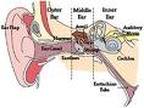
Human Ear
The human ear in transferring sound waves into electrical impulses for the brain to analyses. The ear is divided into three main areas:
- Outer (external) ear
- Middle ear
- Inner ear
Ear
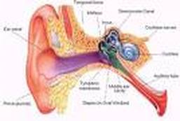
The outer (external) ear
The first step in hearing is the entrance of sound waves into the external auditory canal. The outer ear consists of an outer, funnel-like structure called the auricle(or pinna) and an S-shaped tube, the external auditory meatus. The shapes of the outer ear (the auricle) and the external auditory canal can help amplify and direct the sound. The sound waves reverberate from the sides and end of the external auditory canal, filling it with the continuous vibrations of pressure waves.
The Auricle is what is commonly referred to as the ear. It is composed of irregular plate of elastic cartilage covered with skin, and an occasional hair. Its rim, the helix, is somewhat thicker, and its fleshly, dangling lobule (earlobe) lacks supporting cartilage. The size and shape of the auricle vary considerably from individual to individual, usually being larger in the male than in the female. Though a considerable number of muscle fibres are attached to the auricle, modern man has all but lost the ability to wiggle his ears. The external auditory meatus passes into the temporal bone. Near its opening the tube is guarded by hairs. It is lined with skin that contains numerous modified sweat glands called ceruminous glands, which secrete wax (cerumen). The hairs and wax help to keep relatively large foreign objects, such as insects, from entering the ear.
Sounds generally are created by vibrations of objects that are transmitted through matter in the form of sound waves. For example, the sounds of some musical instruments are produced by vibrating strings or reeds, and the sounds of the voice are created by vibrating vocal folds in the larynx. The auricle of the ear helps to collect sound waves traveling through air and directs them into the auditory meatus. Sound waves entering the external auditory canal eventually hit the tympanic membrane, or eardrum (tympanum = drum), the boundary between the outer and middle ears. The eardrum is a thin, translucent, connective tissue membrane, covered by skin on its external face and by a mucosa internally. It is shaped like a flattened cone, with its apex protruding medially into the middle ear. Sound waves make the eardrum vibrate; the eardrum, in turn, transfers the sound energy to the tiny bones of the middle ear and sets them into vibration.
The middle ear
The middle ear consists of an air-filled space in the temporal bone called the tympanic cavity that separates the external and inner ears, an eardrum or tympanum (tympanic membrane), and three small bones called auditory ossicles. The tympanic membrane is the first structure to respond to sound by vibrating sympathetically with the airborne waves. The tympanic membrane is stretched across the end of the external auditory canal, and air molecules push against the membrane, causing it to vibrate at the same frequency as the sound wave. It has an oval margin and is cone-shaped, with the apex of the cone directed inward. Its cone shape is maintained by the attachment of one of the auditory ossicles (malleus).
The middle ear communicates with the air cells (sinuses) of the mastoid process of the temporal bone and with the nasopharynx via a tube called the pharyngotympanic (auditory) tube (Eustachian tube). Air enters the middle ear through the pharyngotympanic tube to ensure that atmospheric pressure is maintained either side of the tympanic membrane; the equalisation of pressure to vibrate correctly.
The auditory ossicles being the smallest bones in the body are named according to their shape - the malleus (hammer), incus (anvil), and stapes (stirrup). The handle of the malleus is secured to the eardrum, and the base of the stapes fits into the oval window. tiny ligaments suspend the ossicles, and minisynovial joints link them together into a chain that spans the middle ear cavity. The ossicles transmit the vibratory motion of the eardrum to the oval window, which in turn sets the fluids of the inner ear into motion, eventually exciting the hearing receptors.
In addition to transmitting vibrations, the auditory ossicles form a lever system that helps increase (amplify) the force of vibrations as they are passed from the eardrum to the oval window. Since the ossicles transmit vibrations from the relatively large surface of the eardrum to a much smaller area at the oval window, the vibration force becomes concentrated as it travels from the external to the inner ear. As a result of these two factors, the pressure applied by the two stapes at the oval window is about twenty-two times greater than that exerted on the eardrum by sound waves.
The middle ear also contains two small skeletal muscles that are attached to the auditory ossicles. The tensor tympani, is inserted on the medial surface of the malleus, and when it contracts it pulls the bone inward. The other muscle, the stapedius, is attached to the posterior side of the stapes and serves to pull it outward. These muscles are the effectors in the tympanic reflex, which occurs when the ears are assaulted by very loud noises. When the reflex occurs, the muscles contract and the malleus and stapes are moved. As a result, the bridge of ossicles in the middle ear becomes more rigid, and its effectiveness in transmitting vibrations to the inner ear is reduced.
The tympanic reflex is a protective mechanism that reduces pressure from loud sounds that might otherwise damage the hearing receptors. The tensor tympanic muscle also functions to maintain a steady pull on the eardrum. This is important because a loose tympanic membrane would not be able to transmit vibrations effectively to the auditory ossicles.
Eustachian Tube
An eustachian tube (auditory tube) connects each middle ear to the throat. This tube allows air to pass between the tympanic cavity and the outside of the body by way of the throat and mouth. It is important in maintaining equal air pressure on both sides of the eardrum, which is important for normal hearing. The function of the Eustachian tube can be experienced during rapid altitude change. For example, as a person moves from a high altitude to a lower one, the air pressure on the outside of the membrane becomes greater and greater. The eardrum may be pushed inward, out of its normal position, and hearing may be impaired.
When the air pressure difference is great enough, some air may force its way up through the eustachian tube into the middle ear. At the same time, the pressure on both sides of the eardrum is equalized, and the eardrum moves back into its regular position. The person usually hears a popping sound at this moment, and normal hearing is restored. A reverse movement of air ordinarily occurs when a person moves from low altitude into a higher one. The eustachian tube is usually closed by valve like flaps in the throat, which may inhibit air movements into the middle ear. Swallowing, yawning, or chewing may aid in opening the valves, and these actions can hasten the equalisation of air pressure if discomfort is experienced during altitude changes.
The inner ear
The second step in hearing is the transmission of sound energy from the tympanic membrane through the middle-ear cavity to the inner ear. The inner ear consists of a complex system of inter-communicating chambers and tubes called a labyrinth. There are two such structures in each ear - the osseous and membranous labyrinths.
The osseous labyrinth is a bony canal in the temporal bone; the membranous labyrinth lies within the osseous one and has a similar shape. Between the osseous and membranous labyrinths is a fluid, called perilymph, that is secreted by cells in the wall of the bony canal.
The membranous labyrinth contains another fluid, called endolymph, whose composition is slightly different. These fluids serve the dual purpose of cushioning the soft structures and conducting waves from the middle ear to the Organ of Corti, the actual receptor of sound.
The parts of the labyrinths include a cochlea that functions in hearing, and three semicircular canals (anterior, posterior and lateral) that function in providing a sense of equilibrium. A bony chamber, called the vestibule, which is located between the cochlea and the semicircular canals, contains membranous structures (saccule and utricle) that serves both hearing and equilibrium.
Cochlea
The cochlea, as its name suggests, is shaped like the coiled shell of a snail. Inside, it contains a bony core (modiolus) and a thin bony shelf (spiral lamina) that winds around the core like threads of a screw. The shelf divides the bony lanyrinth of the cochlea into upper and lower compartments. The upper compartments, called the scala vestibulli, leads from the oval window to the apex of the spiral. The lower compartment, the scala tympani, extends from the apex of the cochlea to a membrane-covered opening in the wall of the inner ear called the round window. These compartments constitute the bony labyrinth of the cochlea, and they are filled with perilymph. At the apex of the cochlea, the fluids in the chambers can flow together through a small opening (helicotrema).
The membranous labyrinth of the cochlea is represented by the cochlea duct (scala media), which is filled with endolymph. It lies between the two bony compartments and ends as a closed sac at the apex of the cochlea. The cochlear duct is separated from the scala vestibuli by a vestibular membrane (Remissness membrane) and from the scala tympani by a basilar membrane.
The basilar membrane extends from the bony shelf of the cochlea and forms the floor of the cochlear duct. It contains many thousands of stiff, elastic fibres, whose lengths vary becoming progressively longer from the base of the cochlea to its apex. Vibrations entering the perilymph at the oval window travel along the scala vestibuli and pass through the vestibular membrane to enter the endolymph of the cochlear duct, where they cause movements in the basilar membrane.
After passing through the basilar membrane, the sound vibrations enter the perilymph of the scala tympani, and their forces are dissipated to the air in the tympanic cavity by movement of the membrane covering the round window. The Organ of Corti which contains the hearing receptors, is located on the upper surface of the basliar membrane and stretches from the apex to the base of the cochlea. Its receptor cells, which are called hair cells, are arranged in rows and they possess numerous hair like processes that extend into the endolymph of the cochlear duct. As sound vibrations pass through the inner ear, the hairs shear back and forth against the tectorial membrane, and the mechanical deformation of the hairs stimulates the receptor cells. Various receptor cells, however, have slightly different sensitivities to such deformation of the hairs. Thus, a sound that produces a particular frequency of vibration will excite certain receptor cells, while a sound involving another frequency will stimulate a different set of cells.
The cells act very like Neurons in that when it is stimulated appropriately its membrane becomes depolarised, ion channels open, and the membrane becomes more permeable to calcium ions. In the presence of calcium ions, some of the neurotransmitter-containing vesicles in the cytoplasm near its base, fuse with the cell membrane and release neurotransmitter substance into the outside. This neurotransmitter simulates the mends of nearby sensory nerve fibres, and I response they transmit nerve impulses along the cochlear branch of the vestibulo cochlear nerve to the brain. The brain then interprets these nerve impulses, and the hearing process is complete.
Symptoms of Ear Infection in Child
Ear infections often occur in children, at least once before entering the age of 2 years. This is because the connecting pipe between the nose and ear canal contained in children generally have a more horizontal position with the shorter distances, than in adults. Consequently, if there is nasal mucus production in either the flu or allergies, the mucus can be trapped in the channel and into the growth of bacteria. Middle ear infection can cause fever and pain in the ear.
Ear Infection Symptoms in Children
Although ear infections usually occur in children, but in fact can also be experienced by all ages. Common symptom that is felt is the pain in the ear & discomfort. For more details on the ear infection symptoms are:
* Pain in the ear, although it generally occurs but does not always happen.
* Hearing decreases can occur for several days.
* Fever (body temperature increases).
* The child may appear unpleasant taste in the body & puke.
* In infants, usually have not been able to show the pain but it is usually characterized by fussy behavior, fever, frequent crying and refused to eat.
Sometimes it may also occur partly broken ear drum so that infected fluid or pus was flowing out of the ear or commonly known as “congean”. Usually pain will decrease at the ear as it is & a torn eardrum will heal by itself a few weeks later.
Handling of ear infection
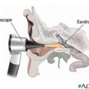
To determine whether there is an ear infection or not, a doctor may look inside your ear using an instrument called the otoscope. Through a doctor’s otoscope will be able to see the condition of the eardrum, which is a thin layer between the outer & inner ear inner ear and blowing air to see if it can be blown eardrum normally. Because in case of an ear infection, the eardrum can not be blown away because it was pressed by the side of pus in it.
Most cases of ear infections can be cured by itself in a few days because normally the immune system can overcome the infection. However, doctors sometimes also provide drugs to help speed up healing, such as:
1. Painkiller
Because ear infections usually cause pain, then pain relievers can be given to patients until the pain healed. Commonly used drugs are paracetamol or ibuprofen. Painkillers may also serve to reduce the fever that accompanies.
2. Antibiotics
Because ear infections can usually heal by itself within 2-3 days then antibiotics are usually not required. Antibiotics may be prescribed by your doctor in case of certain cases, such as:
* Ear infections experienced by children aged <2 years, because the greater the risk of complications in infants.
* Level of ear infections severe enough.
* Ear infections also did not recover within 2-3 days.
* There are complications from an ear infection.
At the time of check to the doctor, a doctor may recommend to see the development of the disease during the next 2-3 days. In a sense it is only for 2-3 days using a painkiller just looking at whether the infection is getting better or not. And if the infection is not improved, then the doctor will prescribe antibiotics.
Ear Care
The ear does not actually need a special thing in her treatment, such teeth should be brushed at least two times a day or a fingernail to be clipped regularly. Care enough with the way the ear is cleaned with soap in the bath or washed with a towel by the shower.
What about ear wax?
Although there was disgusting, ear wax candles work to form a layer that protects the ears. Then after the make, earwax will advance towards the outer ear canal so that it can take a shower washed away during cleaning time. Not recommended for cleaning the ear in a way encouraged to use an object such as cotton buds though, because the dirt can enter into and collected there. The use of cotton buds that are too hard can also irritate the external ear canal. If you want to clean the earwax can use a special fluid that serves to soften the dirt so that it can pass on their own.
