Skeletal System
Human Skeleton
The human skeleton consists of 206 bones. We are actually born with more bones (about 300), but many fuse together as a child grows up. These bones support your body and allow you to move. Bones contain a lot of calcium (an element found in milk, broccoli, and other foods). Bones manufacture blood cells and store important minerals.
The longest bone in our bodies is the femur (thigh bone). The smallest bone is the stirrup bone inside the ear. Each hand has 26 bones in it. Your nose and ears are not made of bone; they are made of cartilage, a flexible substance that is not as hard as bone.
Joints: Bones are connected to other bones at joints. There are many different types of joints, including: fixed joints (such as in the skull, which consists of many bones), hinged joints (such as in the fingers and toes), and ball-and-socket joints (such as the shoulders and hips).
Differences in males and females: Males and females have slightly different skeletons, including a different elbow angle. Males have slightly thicker and longer legs and arms; females have a wider pelvis and a larger space within the pelvis, through which babies travel when they are born.
What is Skeletal System?
The skeletal system (bones and joints), working interdependently with the skeletal muscle system (voluntary or striated muscles), provides basic functions that are essential to life:
A living bone consists of three layers, all honeycombed with nerves and blood vessels: 1) the periosteum, or outside skin of the bone; 2) the hard compact bone, supporting the weight of the body; and 3) spongy bone (bone marrow). Spongy bone occurs at the ends of long bones and is less dense than compact bone. The spongy bone of the femur, humerus, and sternum contains red marrow, producing red blood cells (which carry oxygen), white blood cells (which fight infection), or platelets (that help stop bleeding). Yellow marrow, at the center, is used to store fats. A specialized form of connective tissue, bone consists of both organic components (e.g. collagen) and inorganic minerals (calcium, phosphorus, magnesium, potassium, and sodium). The minerals calcium and phosphorus give bone its hardness, strength, and rigidity to resist compressive forces. The collagen fibers impart flexibility. Magnesium, sodium, potassium, and other trace elements act as "mortar" bonding the calcium and phosphorous. The bone cells themselves are embedded in a mineralized calcium "matrix" and collagen fibers.
Bone continuously remakes itself: New bone is produced and old bone is removed. Osteoblast, the cells responsible for making bone, maintain the balance of calcium in the blood and bone. When this balance is disrupted, as in osteoporosis, the removal of bone exceeds its production, making bone thin and brittle, thus more easily fractured. The intestines, vitamin D, the kidney, parathyroid gland, and sex and adrenal hormones also play important roles in bone/calcium balance. In long bone, illustrated above, growth occurs at the diaphysis side (shaft) of the epiphyseal plate, thus increasing the length of the shaft. Long bone growth stops when the hyaline cartilage stops reproducing itself and fully converts to bone.
A joint, or articulation, is a union of two or more bones. Ligaments attach bone to bone, stabilizing and strengthening joints and determining the range of motion. Cartilage, a gel-like substance high in , proteoglycans, provides protective cushioning. There are three types of cartilage: 1) fibrocartilage (found in intervertebral discs), 2) elastic cartilage (found in the external ear and epiglottis), and 3) hyaline cartilage. Hyaline (or articular) cartilage is the most important cartilage: It serves as the "original" skeleton in the embryo from which bones develop; it spurs growth of long bones; and it lines and protects joints.
- Protection: protects the brain and internal organs
- Support: maintains upright posture
- Blood cell formation: hematopoiesis
- Mineral homeostasis
- Storage: stores fat and minerals.
- Leverage: A lever is a simple machine that magnifies speed of movement or force. The levers are mainly the long bones of the body and the axes (fulcrum) are the joints where the bones meet.
A living bone consists of three layers, all honeycombed with nerves and blood vessels: 1) the periosteum, or outside skin of the bone; 2) the hard compact bone, supporting the weight of the body; and 3) spongy bone (bone marrow). Spongy bone occurs at the ends of long bones and is less dense than compact bone. The spongy bone of the femur, humerus, and sternum contains red marrow, producing red blood cells (which carry oxygen), white blood cells (which fight infection), or platelets (that help stop bleeding). Yellow marrow, at the center, is used to store fats. A specialized form of connective tissue, bone consists of both organic components (e.g. collagen) and inorganic minerals (calcium, phosphorus, magnesium, potassium, and sodium). The minerals calcium and phosphorus give bone its hardness, strength, and rigidity to resist compressive forces. The collagen fibers impart flexibility. Magnesium, sodium, potassium, and other trace elements act as "mortar" bonding the calcium and phosphorous. The bone cells themselves are embedded in a mineralized calcium "matrix" and collagen fibers.
Bone continuously remakes itself: New bone is produced and old bone is removed. Osteoblast, the cells responsible for making bone, maintain the balance of calcium in the blood and bone. When this balance is disrupted, as in osteoporosis, the removal of bone exceeds its production, making bone thin and brittle, thus more easily fractured. The intestines, vitamin D, the kidney, parathyroid gland, and sex and adrenal hormones also play important roles in bone/calcium balance. In long bone, illustrated above, growth occurs at the diaphysis side (shaft) of the epiphyseal plate, thus increasing the length of the shaft. Long bone growth stops when the hyaline cartilage stops reproducing itself and fully converts to bone.
A joint, or articulation, is a union of two or more bones. Ligaments attach bone to bone, stabilizing and strengthening joints and determining the range of motion. Cartilage, a gel-like substance high in , proteoglycans, provides protective cushioning. There are three types of cartilage: 1) fibrocartilage (found in intervertebral discs), 2) elastic cartilage (found in the external ear and epiglottis), and 3) hyaline cartilage. Hyaline (or articular) cartilage is the most important cartilage: It serves as the "original" skeleton in the embryo from which bones develop; it spurs growth of long bones; and it lines and protects joints.
Bone Growth muscle Tissue
 |
 |
General Classifications of Bones
- Long Bones -- "longer than they are wide:" clavicle, humerus, radius, ulna, femur, tibia, fibula, metatarsals, metacarpals. Purpose: provide support and serve as the interconnected set of levers and linkages that allow us to create movement. (formed from hyaline/articular cartilage)
- Short Bones: carpals and tarsals: consist mainly spongy bone covered with a thin layer of compact bone. Purpose: allow movement, provide elasticity, flexibility, & shock absorption.
- Flat Bones: ribs, sternum and scapula. Purpose: protect and provide attachment sites for muscles.
- Irregular Bones: skull, pelvis, and vertebrae. Purposes: support weight, dissipate loads, protect the spinal cord, contribute to movement and provide sites for muscle attachment.
Joints
Joints are classifiied into three groups: 1) immovable (fibrous) joints, e.g. skull bones; 2) slightly movable (cartilagenous) joints, e.g. intervertebral discs; and 3) freely movable (synovial) joints, e.g. limb joints. Synovial joints permit the greatest degree of flexibility and have the ends of bones covered with a connective tissue (synovial membrane) filled with joint (synovial) fluid. A typical synovial joint, seen at right, has four main featues:
- joint capsule - the joint enclosure, reinforced by and strengthened with ligaments
- synovial membrane - a continuous sheet of connective tissue lining the capsule; its cells produce synovial fluid that lubricates the joint and prevents the two cartilage caps on the bones from rubbing together
- synovial fluid - produced by the synovial membrane, the fluid lubricates the joint. In the normal joint, very little fluid (less than 5cc) exists in the cavity.
- hyaline (articular) cartilage - where the bones actually "meet"
Skeleton
The skeleton has two main
parts: the axial skeleton and the
appendicular
skeleton. The axial skeleton consists of the skull, the spine, the
ribs and the sternum (breastbone) and includes 80 bones. The appendicular
skeleton, consisting of 126 bones, includes two limb girdles (the
shoulders and pelvis) and their attached limb bones.
Axial Skeleton (80 bones)
Several structures strengthen the attachments between vertebrae: 1) anterior longitudinal ligaments in front of vertebral bodies and discs; and 2) posterior longitudinal ligaments behind bodies and discs; 3) the compact bone of the disc itself; 4) the interlocking hyaline cartilage surfaces of the neural arch joints; and 5) the ligaments attaching spinous processes to transverse processes.The intervertebral discs provide shock absorption.
Axial Skeleton (80 bones)
- skull - consiting of 1) the cranium (which encloses and protects the brain) and 2) the facial skeleton. The upper teeth are embedded in the maxilla; the lower teeth, in the mandible.
- mandible (jaw) - the only freely movable bone of the skull
- ribs, sternum (breastbone) - comprising the "thorax"/thoracic cage, protecting the heart and lungs
- vertebral column - the "spine"
Several structures strengthen the attachments between vertebrae: 1) anterior longitudinal ligaments in front of vertebral bodies and discs; and 2) posterior longitudinal ligaments behind bodies and discs; 3) the compact bone of the disc itself; 4) the interlocking hyaline cartilage surfaces of the neural arch joints; and 5) the ligaments attaching spinous processes to transverse processes.The intervertebral discs provide shock absorption.
vertebral column vertebra
 |
 |
The orientation of the neural arch joints determines allowable motions: 1) the cervical spine () to rotate, flex forward, flex sideways, and extend backward; 2) the thoracic spine () to rotate; and 3) the lumbar spine () to flex forward, flex sideways, and extend backward. The sacrum () has a dual character, being part of both the vertebral column and pelvis. As such, it transmits the upper body weight to the lower exterminites.
Appendicular skeleton (126 bones, 64 in the shoulders and upper limbs and 62 in the pelvis and lower limbs)
Appendicular skeleton (126 bones, 64 in the shoulders and upper limbs and 62 in the pelvis and lower limbs)
- Upper Extremity - The arms (humerus - upper arm bone) are ultimately attached to the thorax, via synovial joints, at the collarbone (clavicle) and shoulder bone (scapula) (shoulder joint). The scapula is attached to the thoracic cage only by muscles. The elbow joint unites the humerus with the two lower arm bones - the ulna and radius. Three sets of joints connect the radius and ulna to the bones of the palm (metacarpals), via the eight small wrist carpals. Further, the knuckles (metacarpophalangeal, or MCP, joints) connect the metacarpals to the proximal phalanx of the fingers. Each finger has 3 phalanges (proximal, middle, distal), except the thumb which has only two.
- shoulder/ scapula
- arm and forearm, elbow
- hand
- Lower Extremity - The pelvis transmits the upper body weight from the sacrum (at the sacroiliac joint) to the legs. It begins as 3 hip bones (ilium, ischium, and pubis) which fuse together when growth is completed. The hip joint unites the pelvis to the thigh bone (femur); the knee joint, which includes the knee cap (patella), links the femur to the lower leg bones - the tibia and fibula. The ankle joint links the lower leg bones to the talus. The body weight is then transmitted to the heel (calcaneous) and to the balls of the feet via the tarsal and metatarsal foot bones. The toes have a phalangeal structure like the fingers.
- pelvic girdle
- thigh and leg. knee,
- foot/ankle/toe
- Upper Extremity
- Shoulder
- Elbow & Forearm
- Wrist
- Hand
- Spine
- Cervical & Lumbar Spine
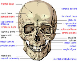
The Skull
The human cranium and the facial bones are the foundation for the soft tissues of the face and head. Thus, much of the visible appearance of the human face depends upon the shapes and qualities of these bones. The cranium is that part of the skull that holds and protects the brain in a large cavity, called the cranial vault. Eight plate-like bones form the human cranium by fitting together at joints called sutures. The most important of these cranial bones for the appearance of the face is the frontal bone, which underlies the top of the face above the eyeballs. The human skull also includes 14 facial bones that form the lower front of the skull and provide the framework for most of the face that is important to psychological research. These 22 skull bones form other, smaller cavities besides the cranial vault, including those for the eyes, the internal ear, the nose, and the mouth. The important facial bones include the jaw bone or mandible, the maxilla or upper jaw, the zygomatic or cheek bone, and the nasal bone.
The shapes and features of the human skull determine much of the static appearances of the face and provide the basis for the features of physiognomy. Forensic pathologists and biologists can reconstruct the superficial appearance of a face merely from the human skull, as in the case of the Kennewick Man. The reconstruction of this skull revealed a facial appearance that indicates he is a descendant of a more ancient migration from Asia than that which brought the ancestors of the Indians (Amerinds), who settled widely in the Americas before the arrival of Europeans.
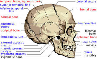
The skull bones are associated with many other features. Processes are areas where the bones have extra tissue to hold muscles and ligaments; lines are grooves in the bone from other developmental processes; foramina are holes in the bones through which nerves and blood vessels pass; sinuses are empty spaces in the bones that make the skull lighter. Some of these features affect the physiognomy of the face due to variations in thickness, size, location, and shape.
The diagrams below show the major external features of the human cranium and the major skull bones. The names in black are facial bones, those in red are cranial bones, and those in blue are features of the bones.
The shapes and features of the human skull determine much of the static appearances of the face and provide the basis for the features of physiognomy. Forensic pathologists and biologists can reconstruct the superficial appearance of a face merely from the human skull, as in the case of the Kennewick Man. The reconstruction of this skull revealed a facial appearance that indicates he is a descendant of a more ancient migration from Asia than that which brought the ancestors of the Indians (Amerinds), who settled widely in the Americas before the arrival of Europeans.
The skull bones are associated with many other features. Processes are areas where the bones have extra tissue to hold muscles and ligaments; lines are grooves in the bone from other developmental processes; foramina are holes in the bones through which nerves and blood vessels pass; sinuses are empty spaces in the bones that make the skull lighter. Some of these features affect the physiognomy of the face due to variations in thickness, size, location, and shape.
The diagrams below show the major external features of the human cranium and the major skull bones. The names in black are facial bones, those in red are cranial bones, and those in blue are features of the bones.
 |
 |
