Muscular System
Muscle cells contain contractile filaments that move past each other and change the size of the cell. They are classified as skeletal, cardiac, or smooth muscles. Their function is to produce force and cause motion. Muscles can cause either locomotion of the organism itself or movement of internal organs. Cardiac and smooth muscle contraction occurs without conscious thought and is necessary for survival.
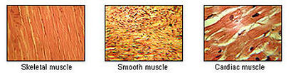
Types Of Muscles
There are three types of muscle:
Skeletal muscle
Smooth muscle
Cardiac muscle
Skeletal muscle
- There are nearly 650 skeletal muscles in the human body!
- Skeletal muscles are attached to the skeleton
- They work in pairs: one muscle moves the bone in one direction and the other moves it back again
- Skeletal muscles are voluntary muscles - in other words we think about what movements we want to make (at least, usually!) and send messages via our nervous system to tell the appropriate muscle(s) to contract.
- Muscle contractions can be short, single contractions or longer ones.
Smooth muscle
- Smooth muscle is found in our internal organs: in our digestive system, our blood vessels, our bladder, our respiratory organs and, in a female, the uterus.
- Smooth muscle can stretch and maintain tension over extended periods
- Smooth muscles are involuntary muscles - in other words we do not have to think about contracting them because they are controlled automatically by the nervous system. It would be pretty inconvenient if we had to think about digesting our food, for example!
Cardiac muscle
- As the name should tell you, cardiac muscle is found only in the heart.
- It can stretch, just like smooth muscle, and contract like skeletal muscle.
- It is a twitch muscle - it only does short single contractions
- Like smooth muscle, cardiac muscle is involuntary. It'd be rather dangerous if it were voluntary - we could stop our heart beating any time we wanted!
|
 |
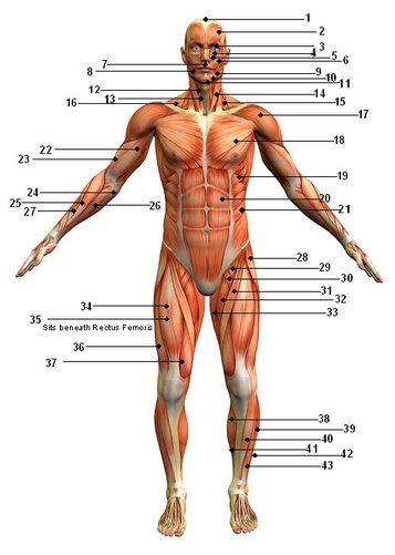
Muscular System-Front
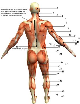
 Muscular System-Back
|
|
|
 |
Movement
- The most well-known function of muscles is movement. Movement when the brain sends electronic signals to the motor neurons on the muscles. Motor neurons are specialized nerves that connect to multiple muscle fibers. As the nerve signals activate the motor neurons, the motor neurons release a neurotransmitter that causes a chemical reaction and the muscles contract.
- Muscles use a tremendous amount of energy to produce movement and one of the by-products of movement is heat. The heat produced from muscle activity helps regulate body temperature and is one of the reasons we shiver when we're cold. Excess heat is carried by the blood to the surface of the skin where the blood is then cooled with sweat evaporation.
- The muscles in our torso keep us upright by maintaining constant tension. They also adjust to compensate when the body is off center. Muscles also have special nerve endings called proprioceptors that help the brain keep track of where our body parts are in relation to each other. It's the proprioceptors that make it possible for you to tilt your head back without falling over, or bring a cup to your lips -- even with your eyes closed.
- The heart is a muscle and pushes blood out into the arteries. Arteries also have a muscle layer that helps push the blood along and provides your pulse. Skeletal muscles also play a role in circulation by assisting with venous return. Unlike arteries, veins have no muscles, no pump and very thin walls. Without assistance from skeletal muscles, the veins have to rely on momentum, existing blood pressure and a system of valves to work against gravity and get blood back to the heart. When muscles contract they act as a pump, by compressing the veins, to assist with blood flow
- Muscles also act as a pump for lymph fluid. Lymph vessels are similar to veins in that they also have no pump, very thin walls and valves to prevent backflow. Lymph vessels run parallel to blood vessels, so the muscle contractions also contribute to lymph flow. The expansion and contraction of the diaphragm, a large circular muscle that assists with breathing, also acts as a major lymph pump in the torso.
- In the absence of movement, venous return and lymph flow slows, causing fluid to pool in the extremities -- this is why feet often swell on long flights.
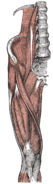
Muscles of Hip
There are several ways of classifying the muscles of the hip:
(1) By location or innervation (ventral an dorsal divisions of the plexus layer);
(2) By development on the basis of their points of insertion (a posterior group in two layers and an anterior group); and
(3) By function (i.e. extensors, flexors, adductors, and abductors). Some hip muscles also act on either the knee joint or on vertebral joints.
Anterior Dorsal Hip Muscles
The anterior dorsal hip muscles are the iliopsoas, a group of two or three muscles with a shared insertion on the lesser trochanter of the femur. The psoas major originates from the last vertebra and along the lumbar spine to stretch down into the pelvis. The iliacus originates on the iliac fossa on the interior side of the pelvis. The two muscles unite to form the iliopsoas muscle which is inserted on the lesser trochanter of the femur. The psoas minor, only present in about 50 per cent of subjects, originates above psoas major to stretch obliquely down to its insertion on the interior side of the major muscle.
Posterior Dorsal Hip Muscles
The posterior dorsal hip muscles are inserted on or directly below the greater trochanter of the femur. The tensor fascia latae, stretching from the anterior superior iliac spine down into the iliotibial tract, presses the head of the femur into the acetabulum but also flexes, rotates medially, and abducts to hip joint. The piriformis originates on the anterior pelvic surface of the sacrum, passes through the greater sciatic foramen, and inserts on the posterior aspect of the tip of the greater trochanter. In a standing posture it is a lateral rotator, but it also assists extending the thigh. The gluteus maximus has its origin between (and around) the iliac crest and the coccyx from where one part radiates into the iliotibial tract and the other stretches down to the gluteal tuberosity under the greater trochanter. The gluteus maximus is primarily an extensor and lateral rotator of the hip joint, and it comes into action when climbing stairs or rising from a sitting to standing posture.
Ventral Hip Muscles
The ventral hip muscles function as lateral rotators and play an important role in the control of the body's balance. Because they are stronger than the medial rotators, in the normal position of the leg, the apex of the foot is pointing outward to achieve better support. The obturator internus originates on the pelvis on the obturator foramen and its membrane, passes through the lesser sciatic foramen, and is inserted on the trochanteric fossa of the femur. "Bent" over the lesser sciatic notch, which acts as a fulcrum, the muscle forms the strongest lateral rotators of the hip together with the gluteus maximus and quadratus femoris. When sitting with the knees flexed it acts as an abductor
Adductor Muscles
The adductor muscles of the thigh are innervated by the obturator nerve, with the exception of pectineus which receives fibers from the femoral nerve, and the adductor magnus which receives fibers from the tibial nerve. The gracilis arises from near the pubic symphysis and is unique among the adductors in that it reaches past the knee to attach on the medial side of the shaft of the tibia, thus acting on two joints. It share its distal insertion with the sartorius and semitendinosus, all three muscles forming the pes anserinus. It is the most medial muscle of the adductors, and with the thigh abducted its origin can be clearly seen arching under the skin. With the knee extended, it adducts the thigh and flexes the hip. The pectineus has its origin on the iliopubic eminence laterally to the gracilis and, rectangular in shape, extends obliquely to attach immediately behind the lesser trochanter and down the pectineal line and the proximal part of the linea aspera on the femur.
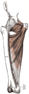
Muscles Of Thigh
The muscles of the thigh can be classified into three groups according to their location: anterior and posterior muscles and the adductors (on the medial side). All adductors (see above) except gracilis insert on the femur and therefore act only on the hip joint. The majority of the thigh muscles, the "true" thigh muscles, are insert on the leg (either the tibia or the fibula) and thus act primarily on the knee joint. Functionally, the extensors lie anteriorly on the thigh and are distinguished from flexors on the posterior side. Even though the sartorius flexes the knee, it is ontogenetically considered an extensor since its displacement is secondarily. Most of the adductors act exclusively on the hip joint, so functionally they qualify as hip muscles.
Anterior Thigh Muscles
The anterior thigh muscles the largest are the four muscles of the quadriceps femoris. The central rectus femoris which is surrounded by the three vasti: The vastus intermedius, medialis, and lateralis. Rectus femoris is attached to the pelvis with two tendons, while the vasti are inserted to the femur. All four muscles unite in a common tendon inserted into the patella from where the patellar ligament extends it down to the tibial tuberosity. Fibers from the medial and lateral vasti form two retinacula that stretch past the patella on either sides down to the condyles of the tibia.
Posterior Thigh Muscles
There are four posterior thigh muscles. The biceps femoris has two heads: The long head has its origin on the ischial tuberosity together with the semitendinosus and acts on two joints. The short head originates from the middle third of the linea aspera on the shaft of the femur and the lateral intermuscular septum of thigh, and acts on only one joint. These two heads unite to form the biceps which inserts on the head of the fibula.
Foot
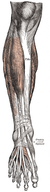
The popliteus (see above) as the single exception, all muscles in the leg are attached to the foot and, based on location, can be classified into an anterior and a posterior group separated from each others by the tibia, the fibula, and the interosseous membrane. In turn, these two groups can be subdivided into subgroups or layers — the anterior group consists of the extensors and the peroneals, and the posterior group of a superficial and a deep layer. Functionally, the muscles of the leg are either extensors, responsible for the dorsiflexion of the foot, or flexors, responsible for the plantar flexion. These muscles can also classified by innervation, muscles supplied by the anterior subdivision of the plexus and those supplied by the posterior subdivision. The leg muscles acting on the foot are called the extrinsic foot muscles whilst the foot muscles located in the foot are called intrinsic.
Dorsiflexion (extension) and plantar flexion occur around the transverse axis running through the ankle joint from the tip of the medial malleolus to the tip of the lateral malleolus. Pronation (eversion) and supination (inversion) occur along the oblique axis of the ankle joint.
Dorsiflexion (extension) and plantar flexion occur around the transverse axis running through the ankle joint from the tip of the medial malleolus to the tip of the lateral malleolus. Pronation (eversion) and supination (inversion) occur along the oblique axis of the ankle joint.
Anterior Foot Muscles
Three of the anterior muscles are extensors. From its origin on the lateral surface of the tibia and the interosseus mebrane, the three-sided belly of the tibialis anterior extends down below the superior and inferior extensor retinacula to its insertion on the plantar side of the medial cuneiform bone and the fifth metatarsal bone. In the non-weight-bearing leg, the anterior tibialis dorsal flexes the foot and lifts the medial edge of the foot. In the weight-bearing leg, it pulls the leg towards the foot. The extensor digitorum longus has a wide origin stretching from the lateral condyle of the tibia down along the anterior side of the fibula, and the interosseus membrane.
Posterior Foot muscles
The posterior muscles three are in the superficial layer. The major plantar flexors, commonly referred to as the triceps surae, are the soleus, which arises on the proximal side of both leg bones, and the gastrocnemius, the two heads of which arises on the distal end of the femur. These muscles unite in a large terminal tendon, the Achilles tendon, which is attached to the posterior tubercle of the calcaneus. The plantaris closely follows the lateral head of the gastrocnemius. Its tendon runs between those of the soleus and gastrocnemius and is embedded in the medial end of the calcaneus tendon.
Intrinsic muscles
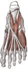
The intrinsic muscles of the foot, muscles whose bellies are located in the foot proper, are either dorsal (top) or plantar (sole). On the dorsal side, two long extrinsic extensor muscles are superficial to the intrinsic muscles, and their tendons form the dorsal aponeurosis of the toes. The short intrinsic extensors and the plantar and dorsal interossei radiates into these aponeuroses. The extensor digitorum brevis and extensor hallucis brevis have a common origin on the anterior side of the calcaneus, from where their tendons extend into the dorsal aponeuroses of digits 1-4. They act to dorsiflex these digits.
The plantar muscles can be subdivided into three groups associated with three regions: those of the big digit, the little digit, and the region between these two. All these muscles are covered by the thick and dense plantar aponeurosis, which, together with two tough septa, form the spaces of the three groups. These muscles and their fatty tissue function as cushions that transmit the weight of the body downward. As a whole, the foot is a functional entity.
The plantar muscles can be subdivided into three groups associated with three regions: those of the big digit, the little digit, and the region between these two. All these muscles are covered by the thick and dense plantar aponeurosis, which, together with two tough septa, form the spaces of the three groups. These muscles and their fatty tissue function as cushions that transmit the weight of the body downward. As a whole, the foot is a functional entity.
